Is a detached retina an eye emergency?
What Is a Detached Retina?
Retinal detachment describes an emergency situation in which a thin layer of tissue (the retina) at the back of the eye pulls away from its normal position.
Retinal detachment separates the retinal cells from the layer of blood vessels that provides oxygen and nourishment to the eye. The longer retinal detachment goes untreated, the greater your risk of permanent vision loss in the affected eye.
Early Signs of a Detached Retina
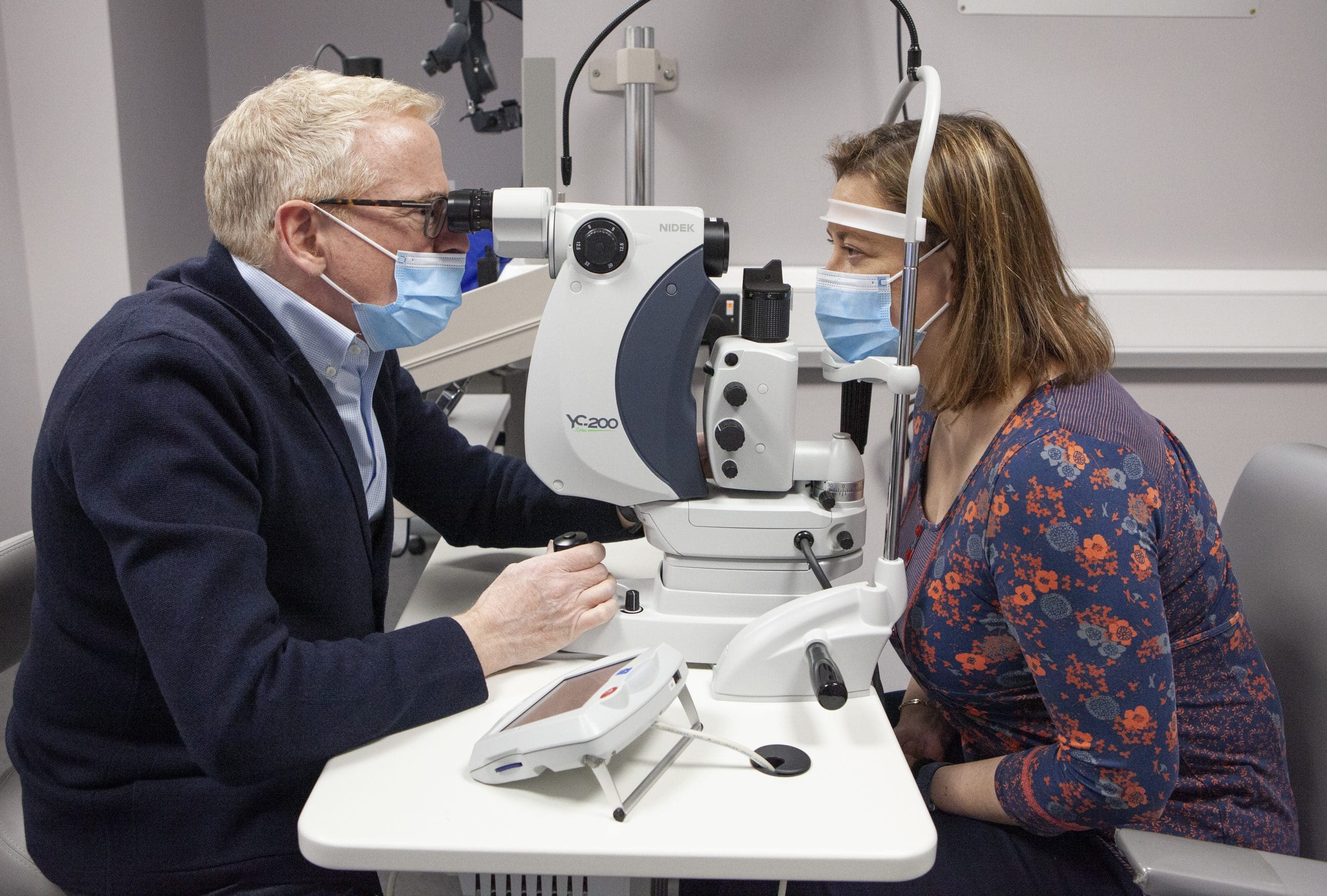
A detached retina has to be examined by an eye doctor straight away. Otherwise, you could lose vision in that eye. Phone Progressive Vision (01 213 5652) immediately if you have any of these symptoms:
- Seeing flashing lights all of a sudden. Some people say this is like seeing stars after being hit in the eye.
- Noticing many new floaters at once. These can look like specks, lines or cobwebs in your field of vision.
- A shadow appearing in your peripheral (side) vision.
- A grey curtain covering part of your field of vision
For out of office hours, please attend the Royal Victoria Eye and Ear Hospital Adelaide Road Dublin – D02 XK51. Phone 01 664 4600
How Do You Get a Detached Retina?
As we get older, the vitreous in our eyes starts to shrink and get thinner. As the eye moves, the vitreous moves around on the retina without causing problems. But sometimes the vitreous may stick to the retina and pull hard enough to tear it. When that happens, fluid can pass through the tear and lift (detach) the retina.
Who Is at Risk for a Retinal Detachment?
You are more likely to have a detached retina if you:
- need glasses to see far away (are nearsighted)
- have had cataract, glaucoma, or other eye surgery
- take glaucoma medications that make the pupil small
- had a serious eye injury
- had a retinal tear or detachment in your other eye
- have family members who had retinal detachment
- have weak areas in your retina (seen by an eye doctor during an exam)
How Is a Detached Retina Treated?
The goal of treatment is to re-attach the retina to the back wall of the eye and seal the tears or holes that caused the retinal detachment.
A detached retina can be treated in a number of ways:
Scleral buckle—In this surgery, a silicone band is placed outside the eye wall to push the wall of the eye closer to the retinal tear in order to close the tear. The tear is treated with a freezing treatment to induce controlled scarring around the tear and permanently seal it. The fluid under the retina is sometimes removed at the time of surgery.
Vitrectomy—In this surgery, three small incisions are made in the white part of the eye and fine instruments are manipulated using an operating microscope to remove the vitreous gel that fills the eye and drain the fluid from under the retina. The surgeon may then use a laser or cryopexy to seal the retinal tears or holes. The eye is filled with a gas or oil bandage to keep the retina dry while it heals.
Pneumatic retinopexy—In this clinic-based procedure, a gas bubble is injected into the eye and the patient maintains a specific head posture to position the gas bubble over the retinal tear. The tear itself is sealed either with a freezing treatment at the time of the procedure, or with laser after the retina is re-attached.
Laser surgery—In certain cases, a retinal detachment can be walled off with laser to prevent the retinal detachment from spreading. This is generally appropriate for small detachments.
Based on the characteristics of the detachment, our retina specialists can determine which treatment is most suitable. In general, retinal detachment repairs succeed in about 9 out of 10 cases, though sometimes more than one procedure is required to successfully put the retina back into place.
What to expect after surgery:
- You might have some discomfort for a few days to weeks after surgery. You will be given pain medicine to help you feel better.
- You need to rest and be less active after surgery for a few weeks. Your eye doctor will tell you when you can exercise, drive or do other things again.
- You will need to wear an eye patch after surgery. Be sure to wear it as long as your eye doctor tells you to.
- If a bubble was put in your eye, you will need to keep your head in one position for a certain length of time, such as 1–2 weeks. Your eye doctor will tell you what that specific head position is. It is very important to follow the directions so your eye heals.
- You might see floaters and flashing lights for a few weeks after surgery. You may also notice the bandage in your eye.
- Your sight should begin to improve about four to six weeks after surgery. It could take months after surgery for your vision to stop changing. Also, your retina may still be healing for a year or more after surgery. How much your vision improves depends on the damage the detachment caused to the cells of the retina.