Retinal vein occlusion

Retinal vein occlusion

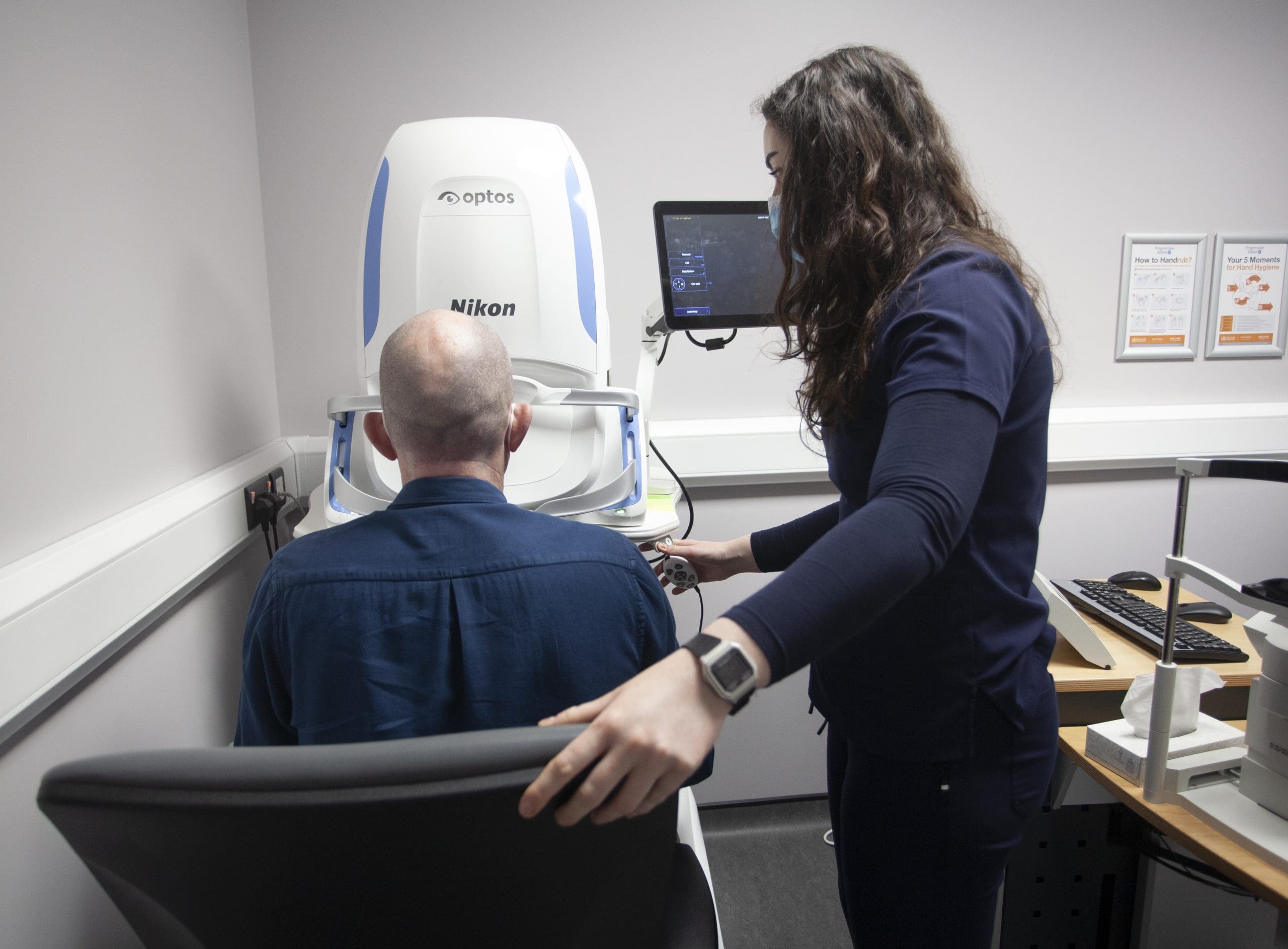
Arteries and veins carry blood throughout your body, including your eyes. The eye’s retina has one main artery and one main vein each of which have 4 main branches.


The arteries change as we get older and can press on the veins slowing down the blood leaving the eye.
As the blood slows, the oxygen the retina needs is reduced and the retina makes chemicals to try and help itself.  The main chemical made is called vascular endothelial growth factor (VEGF) which causes fluid to leak from the blocked veins into the retina causing vision loss. Sometimes VEGF also causes new blood vessels to grow on the retina or in the front of the eye on the iris (coloured part of the eye). Eventually, without blood circulation, nerve cells in the eye can die and you can lose more vision. When the main retinal vein becomes blocked, it is called a central retinal vein occlusion (CRVO). When one of the branches becomes blocked, it’s called a branch retinal vein occlusion (BRVO).

What is Branch Retinal Vein Occlusion (BRVO)?

video from www.aao.org

What is central retinal vein occlusion CRVO ?

video from www.aao.org

What are symptoms of a retinal vein occlusion?

  • The most common symptom of a retinal vein occlusion is vision loss or blurry vision in part or all of one eye.
  • It can happen suddenly or become worse over several hours or days. Sometimes, you can lose all vision suddenly.
  • You may notice floaters. These are dark spots, lines or squiggles in your vision. These are shadows from tiny clumps of blood leaking into the vitreous jelly from new blood vessels on the retina.
  • In some more severe cases of retinal vein occlusion, you may feel pain and pressure in the affected eye

How is a retinal vein occlusion treated?

  • Sometimes steroid medicine may be injected in the eye to help treat the swelling.
  • Sometimes laser can be used in the macula to help oxygen pass from a deeper blood layer in the eye wall into the central part of the retina.
  • This is called argon grid laser  and is usually done after a course of injections but it does not work in every person.
  • If your retinal vein occlusion is very severe and new, abnormal, blood vessels are growing in your eye, you might need a more extensive laser treatment called argon pan-retinal photocoagulation (PRP).
  • This can lower the chance of bleeding in the eye and keeps eye pressure from rising too much.
  • It usually takes a few months after treatment before you notice your vision improving.
  • While most people see some improvement in their vision, some people won’t have any improvement.
  • The severity of the retinal vein occlusion will determine whether your vision will improve with treatment.

Other eye care services.

Age–related macular degeneration (AMD)

A chronic, degenerative condition affecting central vision.

Cataract

A cataract is when your eye’s natural lens core becomes cloudy. This happens as we get older.

Glaucoma

Glaucoma is a disease of the eye where eye pressure is too high, causing damage to your eye’s optic nerve. It usually happens when fluid builds up in the front part of your eye.

Vitreo-retinal surgery

Vitreo-Retinal eye surgery refers to a group of advanced, highly delicate procedures that are done deep inside the eye’s.

General ophthalmology

Light passes through the cornea and lens of each eye.  The cornea and lens focus light on to the back of the eye.

Diabetic eye disease

People with diabetes can have an eye disease called diabetic retinopathy. This is when high blood sugar levels change the blood vessels.

Retinal vein occlusion

Arteries and veins carry blood throughout your body, including your eyes.

Cornea

The cornea is the clear, dome-shaped tissue on the front of your eye that forms the front of the anterior chamber of the eye.

Paediatric ophthalmology

At our specialist paediatric clinic, we work with children in a fun way. This can make eye tests and treatments.